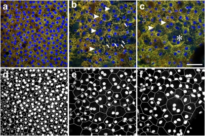
Fig. 3.
Human RPE flat mounts. (A–C) Confocal fluorescence images of RPE flat mounts from equivalent perimacular regions (RPE basal side up). (A) Seventy-six year old (Y) Caucasian males with no history of ocular disease are shown; (B and C) 85 Y Caucasian females with clinically documented dry AMD in an area with multiple binucleate RPE cells (arrowheads in B) and vacuoles (arrows in B) and an area with a trinucleate RPE cell (asterisk in C) is shown. (D–F) A–C are segmented to highlight cell borders and nuclei using DOCAP. (Scale bar: 50 μm.)

