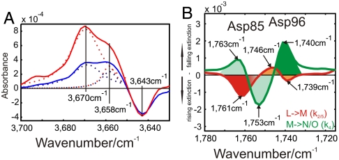
Fig. 4.
FTIR spectroscopy reveals dangling H bonds and Asp96 early changes. (A) M (red) and N (blue) intermediate spectrum differences resulting from time-resolved measurements. In this spectral condition free OH-stretching vibrations (“dangling bonds”) can be observed. The dangling bond of Wat501 at 3,670 cm-1 exhibits a shoulder with kinetically different behavior at 3,658 cm-1. This identifies two neighboring water molecules, because they are influenced by the same mutations (6). The negative band at 3,643 cm-1 reflects the disappearance of the Wat401 dangling bond in the pentameric arrangement (5) (Fig. 1A). (B) Amplitude spectra of the D115N mutant. This mutant was chosen because Asp115 overlaps with the absorbance change of Asp96 in WT (12). We observe a spectral shift of Asp96 from 1,746 to 1,739 cm-1 and Asp85 protonation at 1,761 cm-1 in the L-M transition (11) (red). A spectral shift of Asp85 from 1,763 to 1,753 cm-1 due to changes in H bonding, and Asp96 deprotonation at 1,740 cm-1 in the M-N/O (green) transition (12) are also observed.

