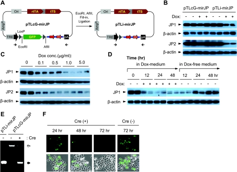
Figure 3.
In vitro assay with Dox-induced knockdown of JP1 and JP2 expression using pTLcG-mirJP system. A) For testing the Dox-induced silencing of JP1 and JP2 expression, the GFP expression cassette was excised from the pTLcG-mirJP plasmid to generate the pTLi-mirJP construct (which mimics Cre-recombination of the pTLcG-mirJP system). B) HEK293 cells in 6-well plates were cotransfected with 1.0 μg of pTLcG-mirJP or pTLi-mirJP together with 0.2 μg of either pCMS-EGFP-mJP1 or pCMS-RFP-mJP2 (41). Dox (1.0 μg/ml) was applied 24 h after transfection. Total cell lysates were prepared at 24 h after addition of Dox, and 10 μg of proteins was analyzed by Western blotting against mouse JP1 or JP2. C) HEK293 cells were cotransfected with 1.0 μg of pTLi-mirJP and 0.2 μg of pCMS-EGFP-mJP1 or pCMS-RFP-mJP2. At 24 h after transfection, cells were treated with indicated concentrations of Dox. Cells were cultured further for 24 h and assayed by Western blotting. D) HEK293 cells cotransfected with 1.0 μg of pTLi-mirJP and 0.2 μg of pCMS-EGFP-mJP1 were treated with Dox (1.0 μg/ml) at 24 h after transfection. After 48 h incubation in the presence of Dox, cells were washed and incubated further in fresh medium as indicated. Expression of JP1 was detected by Western blotting. E) To analyze in vitro Cre recombination, 200 ng of pTLcG-mirJP was incubated with Cre enzyme (1 U) at 37°C for 30 min, followed by heat inactivation of Cre at 70°C for 10 min. Plasmids were purified from the reaction mixture, and the recombined plasmids were analyzed by PCR, using 2 primers flanking the GFP expression cassette. Open arrow indicates unrecombined product (2030 bp) containing the GFP expression cassette; solid arrow indicates recombined product (470 bp). F) HEK293 cells were transfected with 0.2 μg of pTLcG-mirJP with or without 1.0 μg of pTurbo-Cre. From 24 h after transfection, cells were observed by fluorescence microscopy to detect GFP expression.

