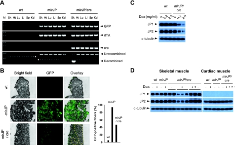
Figure 5.
Muscle-specific knockdown of JP1 and JP2 expression using the mirJP/Cre transgenic mice. pTLcG-mirJP transgenic (mirJP) mice were mated with HSA-Cre79 transgenic (cre) mice to generate offspring carrying both transgenes (mirJP/Cre). Eight-week-old F2-mirJP/cre mice were examined to detect Cre-mediated DNA recombination (A, B) and Dox-dependent JP knockdown (C, D). A) Genomic PCR was performed in various tissues of wild-type (wt), pTLcG-mirJP (mirJP), and pTLcG-mirJP/HSA-cre (mirJP/cre) transgenic mice. Cre-mediated recombination was determined by detection of two different sizes of PCR products, 2030 bp for unrecombined and 470 bp for recombined products. M, 100-bp DNA ladder marker; Sk, skeletal muscle; Ht, heart; Lu, lung; Li, liver; Sp, spleen; Kd, kidney. Asterisks indicate nonspecific PCR products. B) Gastrocnemious muscles prepared from the mice were cross-sectioned (5 μm thickness) and examined under fluorescence microscopy to visualize the expression pattern of GFP. Area enclosed by dashed line corresponds to fibers showing negative or very weak GFP fluorescence on the section. GFP-positive fibers were counted from the same sections (right panel). C) Examination of Dox-dependent reduction of the JP expression in skeletal muscle. Eight-week-old wt and mirJP/cre mice were supplied with water containing 0.2 or 2.0 mg/ml of Dox for 1 wk. Gastrocnemious muscles were isolated, and expression levels of JP1 and JP2 were examined by Western blotting. D) Reversible control of Dox-inducible knockdown of JPs in skeletal muscle. Eight-week-old wt and transgenic mice were provided with water either with or without Dox (2 mg/ml) for 2 wk. One cohort of Dox-treated mirJP/cre mice was sacrificed (Dox + group), and the other cohort was supplied with Dox-free water for 4 wk to inactivate Tet-on system for shRNA-mirJP expression and recover expression level of JP1 and JP2 (Dox +→− group). Gastrocnemious and cardiac muscles were prepared and examined by Western blotting for JP-1 and JP-2. Each lane shows Western blotting for the muscle isolated from different mice.

