Figure 1.
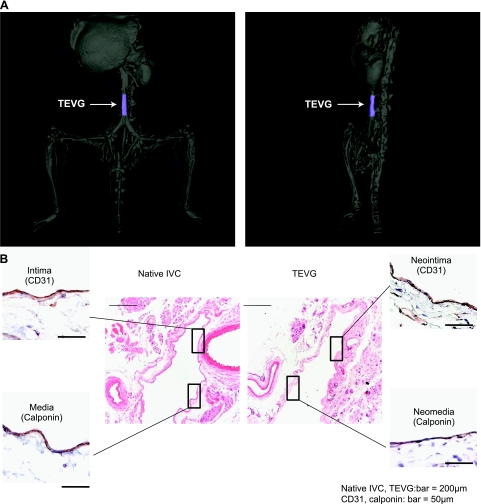
A) Representative CT angiogram of a TEVG IVC interposition graft 6 mo after implantation. TEVG is colored blue. B) Representative photomicrographs of immunohistochemical characterization of the native IVC compared with the TEVG 6 mo after implantation (hematoxylin and eosin, ×100; CD31 and Cal, ×400).

