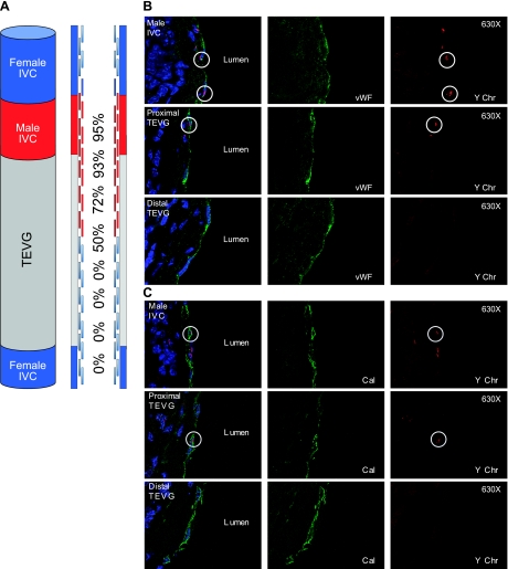
Figure 5.
A) Schematic demonstrating composite TEVG (n=8) created by anastomosis of a syngeneic male IVC with a TEVG, implanted into a female host and harvested at 6 mo after implantation, and the percentage of cells with Y-chromosome as a function of distance from the male IVC. A) Confocal microscopic images demonstrating colocalization of endothelial cells (vWF) and Y chromosome (Y-chromosome FISH; ×400). C) Photomicrographs demonstrating colocalization of smooth muscle cells (calponin) and Y chromosome (Y-chromosome FISH; ×400).

