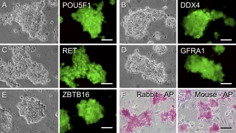
Figure 3.
Characterization of rabbit clump-forming cells. Continuously proliferating cell clumps were cultured on C166 feeder cells in serum-free medium supplemented with GDNF, GFRA1, and FGF2. A–E) Phase contrast (left panels) and immunofluorescent images (right) of clump-forming cells stained with antibodies against POU5F1 (A), DDX4 (B), RET (C), GFRA1 (D), and ZBTB16 (E), which are expressed in mouse SSCs, followed by staining with Alexa Fluor 488-conjugated appropriate secondary antibodies. Rabbit clump-forming cells expressed the SSC markers. F) AP activity of rabbit clump-forming cells and cultured mouse SSCs. Scale bars = 50 μm (A–E); 100 μm (F).

