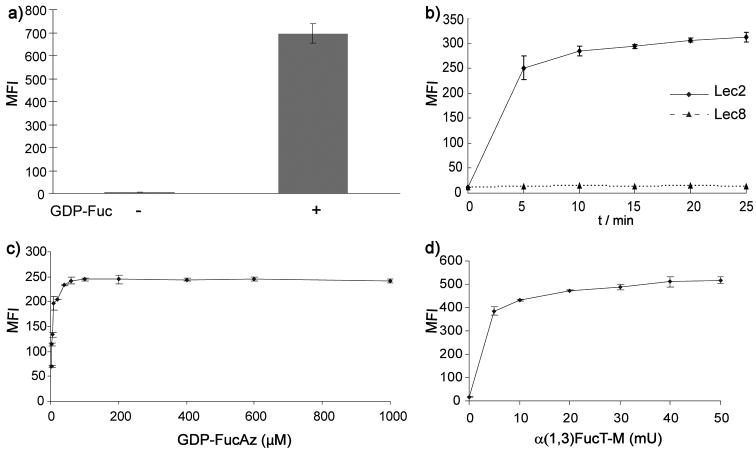
Figure 2.
Detection of cell surface glycans with LacNAc using flow cytometry. a) Lec2 CHO cells were treated with or without GDP-Fuc (500μM) in the presence of α(1,3)FucT-M for 10 min, and probed with PE-conjugated anti-LeX IgM. b) Time-dependent labeling of cell surface glycans with LacNAc. Lec2 and Lec8 CHO cells were treated with GDP-FucAz (500 μM) and α(1,3)FucT-M (30 mU) for 0–25 min. The labeled cells were then reacted with BARAC-biotin (10 μM) for 10 min and then probed with streptavidin-Alexa Fluor 488. c) and d) dose-dependent labeling of cell surface glycans with LacNAc. c) 30 mU α(1,3)FucT-M, labeling time for 10 min. d) 500 μM GDP-FucAz, labeling time for 10 min. Error bars represent the standard deviation of three replicates in one experiment.

