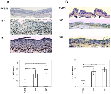
Figure 3. Analysis of cellular proliferation in the ear and dorsal skin of wild-type and transgenic mice.
(A and B, top panels) Representative pictures of Ki-67 immunostained sections of paraffin-embedded ear skin and dorsal skin from wild-type (FVB/N) and Tg animals (lines 183 and 187). (A and B, lower panels) Quantification of Ki-67-positive cells (brownish signal) in wild-type and Tg epidermis was done by counting 400 hematoxylin-stained cells under 40× magnification in 4 different fields of epidermis. Differences between the Ki-67-positive cells in the HPV38 E6/E7 Tg mice (lines 183 and 187) versus the FVB/N mice were statistically significant as determined by Student's t-test with Welch correction for unequal standard deviation.

