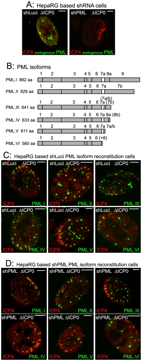
Figure 1. The major nuclear isoforms of PML and their recruitment to sites associated with HSV-1 genomes.
A. Typical examples of recruitment of endogenous PML (green) to sites associated with HSV-1 genomes (ICP4: red) in cells at the edges of ICP0 null mutant (ΔICP0) plaques in control and PML depleted cells. B. PML isoforms I to VI, noting the included exons and the translated product length. The bracketed exons indicate the use of alternative reading frames for these sequences. (+8) at the end of PML VI indicates an additional 8 residues following exon 6 as a result of alternative splicing that deletes exon 7a. Adapted from [23]. C and D. Recruitment of EYFP-PML isoforms in control (panel C) and PML depleted cells (panel D). Scale bars indicate 5 µm. Analysis of the localization of these proteins in uninfected cells and their expression levels is presented elsewhere [23].

