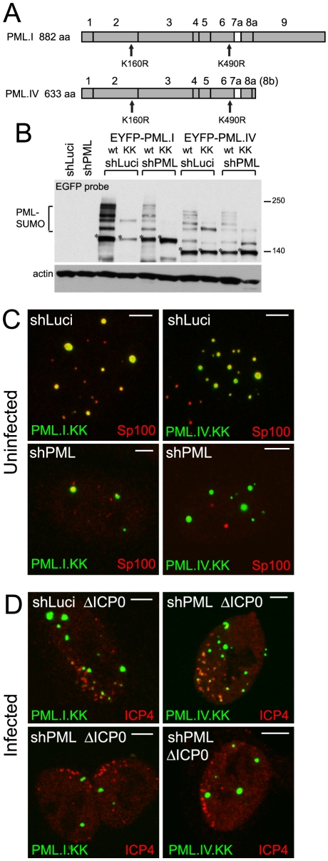
Figure 3. SUMO modification mutants of PML are defective in recruitment to sites associated with HSV-1 genomes.
A. Maps of PML isoforms I and IV and derivatives with lysine to arginine substitutions at residues K160 and K490. B. Western blot of these proteins in control and PML depleted cells, using an anti-EGFP antibody. The major unmodified bands of the EYFP-PML proteins are indicated by asterisks. C. Localization of the EYFP-PML proteins (green) and endogenous Sp100 (red) in uninfected control and PML depleted cells. D. Typical assays of recruitment of these PML proteins (green) to sites associated with HSV-1 genomes (ICP4; red) in ICP0 null mutant (ΔICP0) infected control and PML depleted cells. Scale bars indicate 5 µm.

