
Figure 9. Recruitment of SUMO family members and PIAS2β to HSV-1 induced foci.
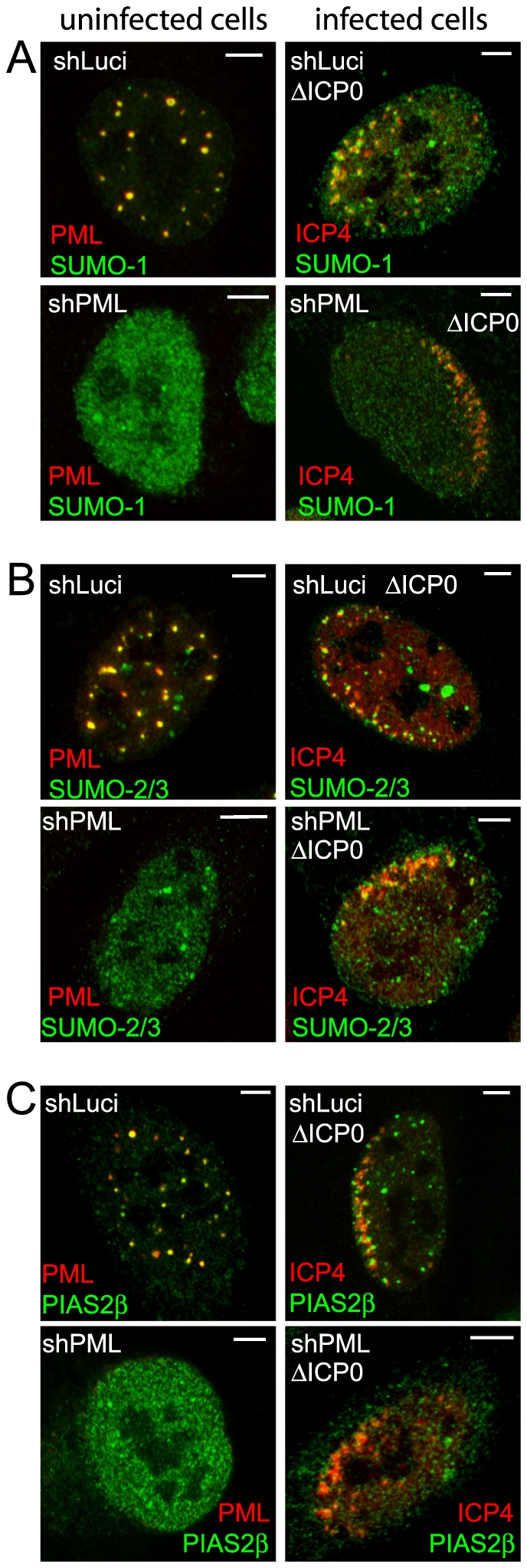
Left-hand images show uninfected cells and the co-localization of SUMO-1 (A), SUMO-2/3 (B) and PIAS2β (C) (green) with PML (red) in control (upper rows of each block of 4 images) and PML depleted (low rows of each block of 4 images) HepaRG cells. Right-hand images show typical examples of recruitment of the indicated proteins to sites associated with HSV-1 genomes (ICP4; red) in cells at the edges of ICP0 null mutant (ΔICP0) plaques in control and PML depleted HepaRG cells. Scale bars indicate 5 µm.