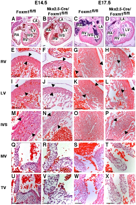
Figure 3. Myocardial thinning and cardiomyocyte disarray in Nkx2.5-Cre/Foxm1fl/fl mice.
Microtome sections of paraffin-embedded hearts were prepared from Nkx2.5-Cre/Foxm1fl/fl and control Foxm1fl/fl mice at E14.5 and E17.5 and stained with hematoxylin and eosin (H&E). H&E staining showed that hearts from Nkx2.5-Cre/Foxm1fl/fl embryos possessed all four chambers and were similar in size to littermate control hearts (A–D). However, there was thinning of the muscular wall of the right ventricle (RV) (E–H), left ventricle (LV) (I–L), and interventricular septum (IVS) (M–P) in addition to disorganization of the musculature. There was no difference in the size of the leaflets in either the mitral (MV) (Q–T) or tricuspid valves (TV) (U–X). Arrowheads indicate area where measurements of thickness were made. Scale bars represent 100 µm in A–B and E–P, 200 µm in C–D and 50 µm in Q–X.

