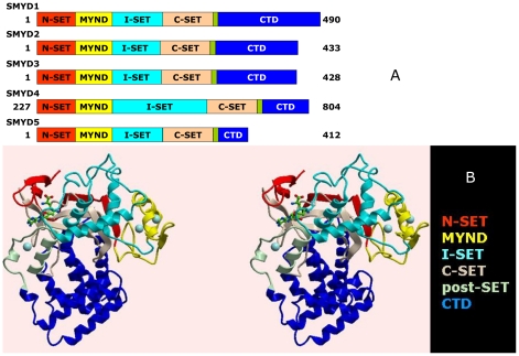
Figure 1. Structure of SMYD3 and its paralogs.
(A) Linear representation of domain structures of SMYDs1–5. The split SET domain is shown in red (N-SET) and tan (C-SET); the MYND domain is represented in yellow and the cysteine-rich post-SET domain is displayed in pale green. Starting and ending amino acids are indicated. (B) Ribbon representation of the structure of SMYD3-Sinefungin at 1.85Å resolution in cross-eye stereo. The SET domain of SMYD3 is split into the N-SET (red) and the C-SET (tan) by an intervening MYND domain (yellow) and a Rubisco-LSMT-like I-SET region (cyan). The post-SET motif (pale green) precedes a long (∼150 residue) C-terminal domain (CTD, blue). Positions of Sinefungin (green carbons) and zinc atoms (spheres) are indicated.

