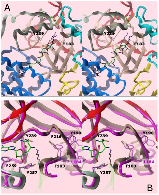
Figure 5. Model of the SMYD3-Sinefungin active site with the H3K4me1 peptide [41] in cross-eye stereo.
The peptide, colored black, and with backbone traced in green ribbon, was taken from an overlay with the SET9 ternary structure. (A) Ribbon representation of the ternary complex. Substrate methyl donor and peptide are indicated as green wire bonds and ribbons, respectively. Domain colors for SMYD3 correspond to those in Fig. 1. The aromatic cage residues (Y239, F183) at the end of the lysine channel are shown explicitly. (B) Overlay of SMYD1 (magenta, PDB accession #3N71) and SMYD3 (colored by domain) proteins in the ternary model. Numbering of residues is for SMYD3.

