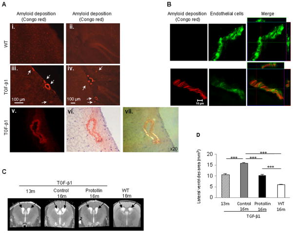
Fig. 1.
Nasal Protollin reduces brain damage in TGF-β1 mice. (A) Congo red staining in the brains of 16-month-old TGF-β1 Tg mice (A, iii–vi) and age-matched non-transgenic littermates (WT) (A, i–ii). Vascular amyloid (Congo red) is apparent in the cortex (A,iii, scale bar: 100 μm ) and hippocampus (A,iv, scale bar: 100 μm) of a 16-month-old TGF-β1 Tg mice. Fluorescence image (v) and brightfield microscopy image (vi), after Congo red staining. Scale bar: 50 μm. (B) Colocalization (right) of vascular amyloid (Congo red, left) and endothelial cells (EC) (CD31 marker, green, middle) in the brain of 16-month-old TGF-β1 Tg mice compared to WT mice. Scale bar: 10 μm. (C) T2-weighted representative images and (D) analysis of the lateral ventricle area (mm2) of TGF-β1 mice brain with (n=7 mice) and without Protollin treatment (n=5 mice). Arrows point to lateral ventricles (**P<0.01, *** P<0.001).

