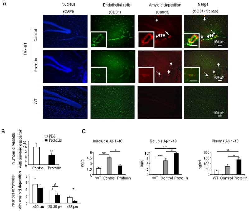
Fig. 3.

TGF-β1 mice treated with Protollin showed reduction in vascular amyloid. (A) Double-label immunofluorescent staining of vascular amyloid (Congo red) and endothelial cells (CD31) in brain sections of control mice compared to Protollin treated mice and WT mice. Scale bar: 100 μm. (B) Quantified analysis of the number of vessels containing amyloid deposition in brain section of control and Protollin treated group (upper panel) and according to the size of affected vessels (lower panel). Comparisons were made using unpaired t tests (**P<0.03, #P=0.05, *P<0.04; n=5 mice/ group). (C) Brain insoluble, soluble and plasma mouse Aβ (1–40) levels were measured by ELISA (*P<0.05, **P<0.01, ***P<0.001; n=5 mice/ group).
