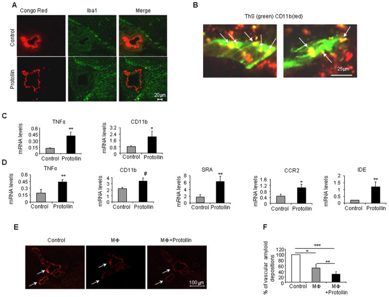
Fig. 5.

Cerebrovascular amyloid clearance in Protollin-treated mice is mediated by activated macrophages. (A) Activated perivascular macrophage cells (Iba1, positive) colocalized with a reduction of vascular amyloid deposition (Congo red) in the brains of Protollin-treated mice vs. control mice. Scale bar: 20 μm. (B) Confocal image of colocalization of CD11b positive cells (red) with vascular amyloid (ThS green) in Protollin treated mice. Scale bar: 50 μm. (C) Elevation of monocyte mRNA expression markers TNF-α (**P<0.02) and CD11b (*P<0.05) in the blood of Protollin-treated mice compared to the control mice (n=6 mice/group). (D) Elevation of macrophage activation markers from Protollin-treated mice compared to control: SRA (**P<0.02), TNFα (**P<0.02), IDE (**P<0.02), CD11b (#P<0.04), CCR2 (*P<0.05 from 16 months old immunized TGF-β1 Tg mice treated with Protollin (n=7) or PBS (n=6). (E) Representative figures and (F) analysis of reduction of cerebrovascular amyloid (Congo red) following incubation for 4 days at 37° C with Macrophage cell line (RAW264.7) with (right panel) and without (middle panel) 0.1 μg/ml Protollin ( *P<0.05, **P<0.0003, ***P<0.0001, n = 7).
