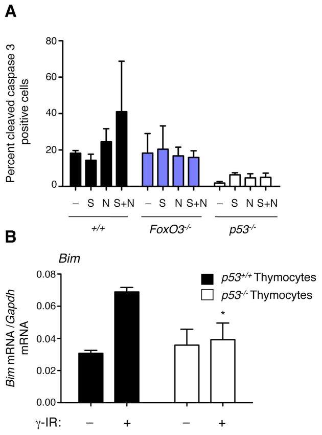
Figure 7. FoxO3 plays a role in p53-dependent apoptosis.
(A) Percent cleaved caspase 3-positive cells in E1A-transformed MEFs (FoxO3+/+p53+/+ (+/+), FoxO3−/−, and p53−/−) in the absence of treatment (−) or in response to Nutlin (N), serum starvation (S), and Nutlin + serum starvation (N+S). Mean +/− SEM of three independent experiments. (B) Real time quantitative PCR analysis of Bim mRNA levels in p53+/+ and p53−/− thymocytes, 3 hours after γ irradiation (γ IR, 10 Gy). Mean +/− SEM of two independent experiments conducted in triplicate on samples from 3–5 mice per genotype. * p<0.05 between p53+/+ and p53−/− thymocytes at a given time point, two-way ANOVA with Bonferroni post-test.

