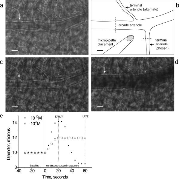
Figure 1.
Representative images of the arcade arteriole-terminal arteriole junction with micropipette placement for curcumin administration (schematic, B) in the basal control state (A), constricted with phenylephrine (C), and dilated with adenosine (D). Dashed lines in (B) indicate typical locations for diameter measurements. White arrow points to a bulge, which is a vascular smooth muscle cell in cross section; especially in (C) the cells are seen as a `string of pearls' along the vascular wall. With dilation (D) the cells flattens and are not apparent. (E) Time course of the diameter change in response to continuous curcumin application (micropipette) in one terminal arteriole. Low dose curcumin induced a sustained vasodilation, peaking by 20 seconds. Higher dose curcumin initiated dilation, peaking by 20 seconds, followed by a secondary constriction, peaking by 60 seconds. Data for concentration response analysis was thus taken at both the EARLY (20 second) and LATE (60 second) time periods. Scale bar in A,B,C,D is 20 microns.

