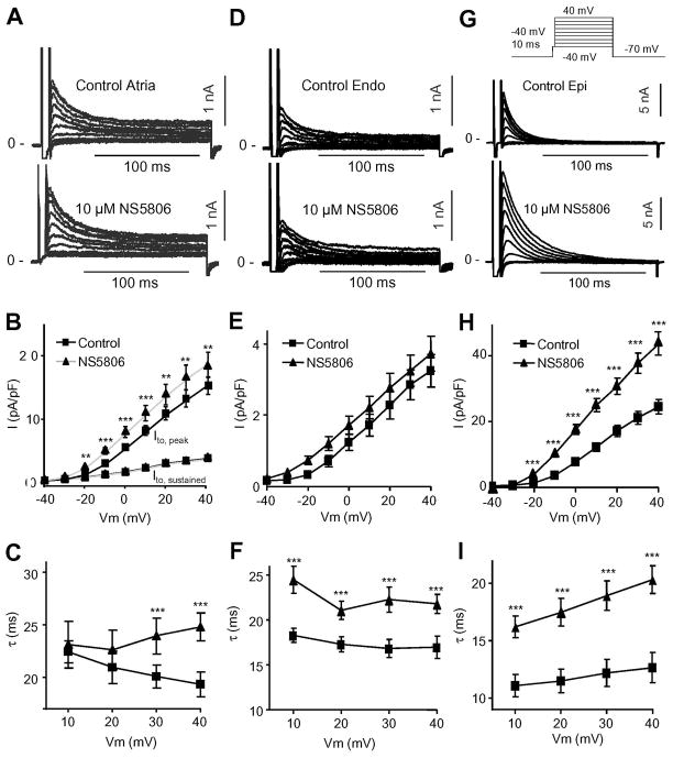
Figure 2.
Ito in canine atrial and ventricular cells in absence and presence of NS5806 (10 μM). Currents were activated by the voltage clamp protocol (top of figure) and Cd2+ was present to block ICaL. Panel A) Representative Ito traces recorded from canine atrial cells. Panel B) Mean I–V relation for atrial peak Ito as well as for the sustained component Panel C) Ito decay as a function of voltage under control (n=14). Panel D) Representative Ito traces recorded from canine LV Endo cells. Panel E) Mean I–V relation for peak Ito. Panel F) Ito decay as a function of voltage (n=9). Panel G) Representative Ito traces recorded from canine LV Epi cells. Panel H) Mean I–V relation for peak Ito. Panel I) Ito decay as a function of voltage (n=10).

