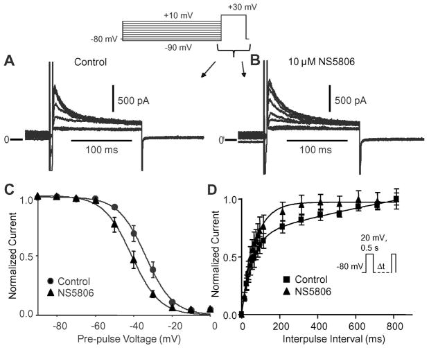
Figure 3.
Voltage dependence of inactivation of atrial Ito. Representative traces recorded under control conditions (Panel A) and after application of 10 μM NS5806 (Panel B) showing voltage dependence of inactivation of Ito. Panel C) The currents were normalized to max-current and plotted as a function of the conditioning pulse and Boltzmann curves were fit to the data. In the absence of drug, the mid-inactivation voltage was V1/2=−33.8±0.46 mV and the slope factor k=−7.15±0.40. In presence of NS5806 V1/2 was −41.1±0.50 mV and k=−7.35±0.42 (n=8). Panel D) Time-dependence of recovery from inactivation of atrial Ito. Currents were elicited by the depicted protocol and normalized recovered currents were plotted as a function of the interpulse interval (n=6).

