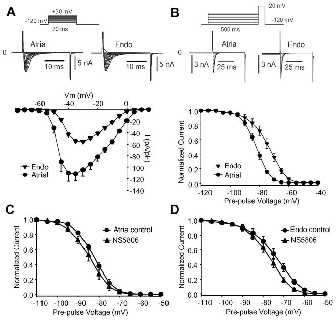
Figure 4.
INa recorded from canine atrial cells or endocardial cells. Cd2+ was present to block ICaL. Panel A) Representative INa traces recorded from atrial (n=8) and Endo cells (n=9) and corresponding mean I–V relation for peak current is shown. Panel B) Representative steady-state inactivation recordings from an atrial or Endo cell. The peak-current following a 500 ms prepulse was normalized to the maximum current and plotted against the conditioning potential to obtain the availability of channels. A Boltzmann equation was fit to the data resulting in a mid-inactivation (V½) of −82.6±0.12 mV (n=8) for atrial and −73.9±0.27 mV (n=9) for Endo I Na. Panel C) Steady-state inactivation for INa recorded from atrial cells in absence and presence of 10 μM NS5806. V½= −82.6±0.1 mV in control and −85.1±0.1 mV in presence of NS5806 (n=8). Panel D) Steady-state inactivation for INa recorded from isolated LV Endo cells in absence and presence of 10 μM NS5806. V½= −73.9±0.27 mV in control and −77.3±0.21 mV in presence of NS5806 (n=9).

