
Figure 2. Voxel-wise edge displacement results.
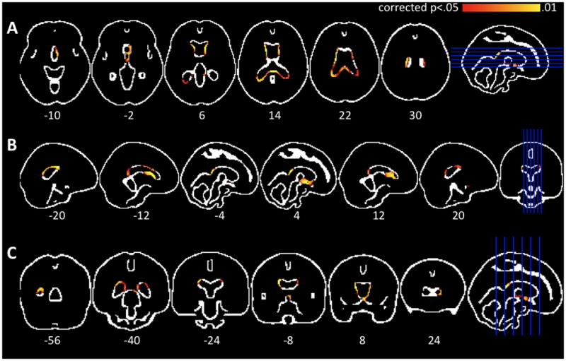
Axial(A), saggital(B), and coronal (C) images of the MNI152 edge image overlaid with areas of significant differences between close-to-onset individuals and controls (p<.05, corrected over the edge image). There is significant periventricular edge displacement, consistent with progressive basal ganglia atrophyin premanifest Huntington’s disease.