
Figure 5.
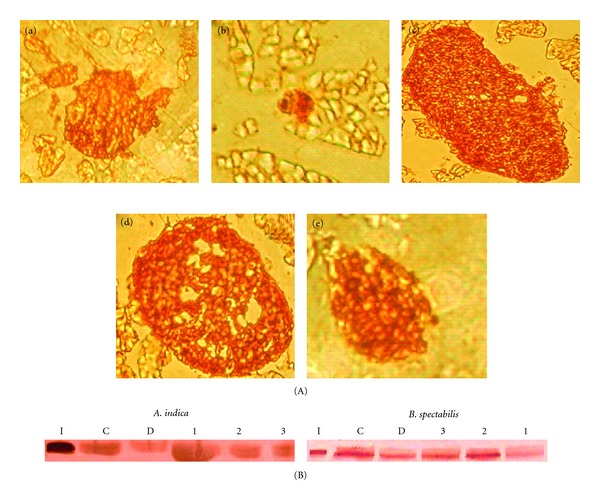
(A) Immuno-histochemical analysis of the mouse pancreas developed with anti-insulin antibody. (a) Normal, (b) Diabetic, (c) A. indica chloroform extract treated, (d) B. spectabilis aqueous extract treated, (e) B. spectabilis methanolic extract treated with magnification of 480x, and (B) immunoblot analysis of pancreatic protein probed with anti-insulin antibody after treatment with (I) Human recombinant insulin, (C) control, and (D) diabetic (1) chloroform (2) methanolic (3) aqueous. The data are measured by densitometry in ODU/mm2.