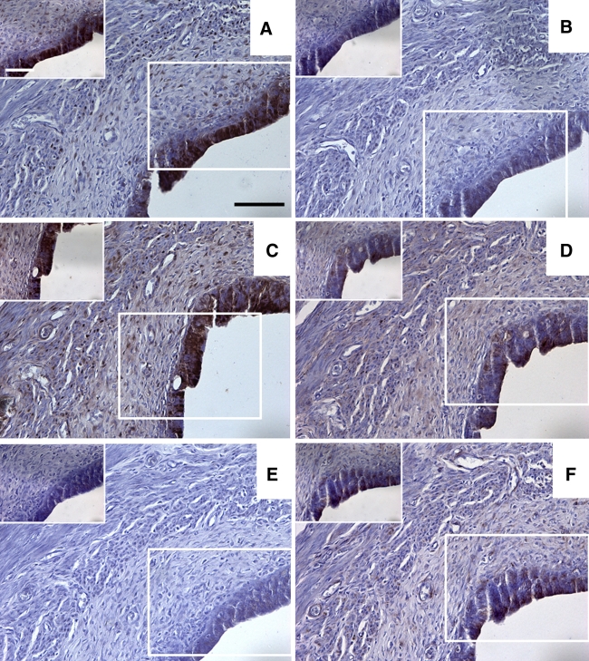
Fig. 3.
a–f Sections taken from a representative rat showing the results for immunohistochemical staining in the uterus for pimonidazole, HIF1α, HIF2α, GLUT1, GLUT4 and CAIX, respectively. The boxed diagrams in a–f show a higher magnification detail for each stain. Scale bars 100 μm (a–f) or 30 μm (detail in a–f)

