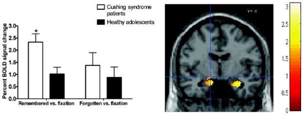
Figure 2.
Mean BOLD signal differences in the left amygdala (MNI â??14, â??6, â??10Â mm) for contrasts remembered versus fixation and forgotten versus fixation in Cushing syndrome patients and healthy adolescents. Adolescents with Cushing syndrome showed significantly higher activation when compared to healthy adolescents (p < .05). Error bars represent standard error of the mean.
The regions of interest (ROI) analyses for the left amygdala and right anterior hippocampus. The statistical threshold is set at p < .05 after a small volume correction of the ROI.

