
Figure 1.
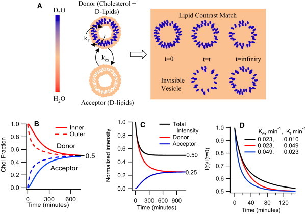
(A) Schematic of experimental protocol: Donor and acceptor vesicles made of dPOPC with a matrix (solvent) completely matched to the lipids rendering them invisible to neutrons. At t = 0, only the donor vesicles are visible as they contain cholesterol, the only visible component in the mixture. As cholesterol moves from donor to acceptor vesicle, the acceptor vesicles gradually begin to appear. (B) Cholesterol fractions in the inner and outer monolayers of the donor and acceptor vesicles as a function of time assuming cholesterol exchange follows first order kinetics and Kex and Kf = 0.01 min−1. (C) Contribution to the normalized intensity from the donor and acceptor vesicles as a function of time assuming the rates above. (D) Plot of normalized intensity decay curves illustrating the relative effects of Kex and Kf on the decay curves.