
Figure 2.
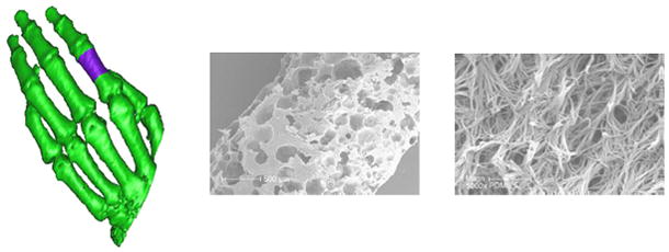
Conversion of CT images into micro- and nanostructure controlled PLA scaffolds. A CT image of a hand (left) with a non-traditional defect (shown in purple) is converted into a wax mold which can be filled with PLA to create a scaffold with controllable pore size on the micro scale (center) and fiber size on the nano scale (right). (reprinted from 65 with permission from Elsevier.)