
Figure 7.
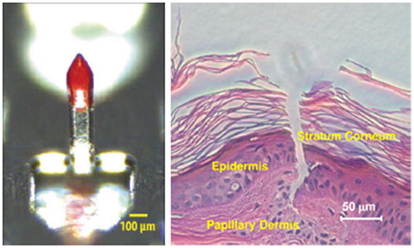
Optical microscopy image of a polyphosphazene coated metallic microneedle (left) and histological section of porcine skin after coated microneedle insertion (right). (reprinted from 310)
Official websites use .gov
A
.gov website belongs to an official
government organization in the United States.
Secure .gov websites use HTTPS
A lock (
) or https:// means you've safely
connected to the .gov website. Share sensitive
information only on official, secure websites.
Optical microscopy image of a polyphosphazene coated metallic microneedle (left) and histological section of porcine skin after coated microneedle insertion (right). (reprinted from 310)