
Figure 8.
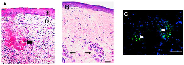
Photomicrographs of keratinocyte-fibroblast co-culture engineered skin tissue with the addition of collagen microsphere supported sweat gland cell constructs. (A) Hemotoxylin and Eosin (H&E) staining after two weeks of in vitro co-cultivation showed differentiated tissue layers (E: epithelium and D: dermis) with a bud-like structure (black arrow) where sweat gland constructs were loaded. (B) H&E staining of six week post-implantation in vivo skin tissue showed the continued presence of bud-like structures in the dermis layer (black arrow). (C) Fluorescence microscope observation showed DiO-positive cells (green) confirming the presence of still viable sweat glands. (reprinted from 485 with permission from Elsevier.)