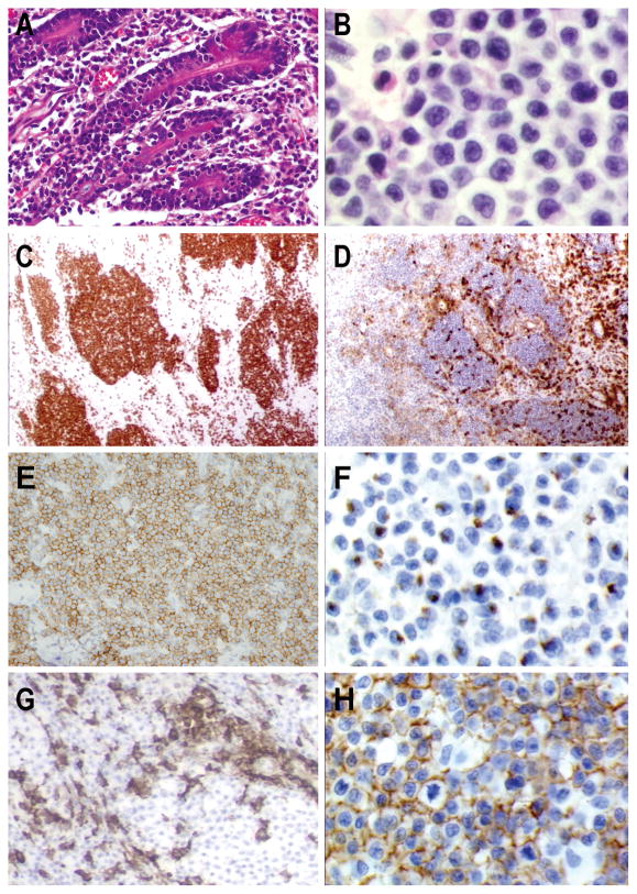
Figure 3. T-cell lymphoma of γδ origin with intestinal involvement. Type II variant of enteropathy-associated T-cell lymphoma.
A) The tumor surround mucosal crypts with intraepithelial involvement (H&EX20) B) Monotonous lymphoid infiltrate of small medium sized tumor cells (H&E X60) The atypical lymphocytes are C) CD8 positive (X10), D) CD5 negative (X10) and E) CD56 positive (X40) F) Neoplastic lymphocytes have an activated cytotoxic phenotype with granzyme-B expression (X60) G) Reactive lymphocytes are positive for TCRβ that is absent in tumor cells (X40) H) Homogenous TCRδ expression is observed in the tumor (X60)

