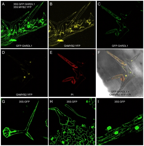
Figure 7. Subcellular localization of GFP:GhRDL1 and GhMYB2:YFP.
(A–B) Localization of GFP:GhRDL1 (A) and YFP:GhMYB2 (B) in silique trichomes. (C–F) Silique trichomes in the try transgenic plants overexpressing GhRDL1 and GhMYB2 stained with GFP (C), YFP (D), PI (E, red), and merged (F). (G–I) Diffusion of GFP in leaf trichomes (G), epidermal cells (H), and siliques (I) in 35S:GFP transgenic plants. GFP donuts in (I) indicate guard cells of stomata.

