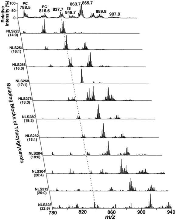
Figure 2. Representative 2D MS analyses of TAG species in a human plasma lipid extract.
Neutral loss scans (NLS) of all naturally-occurring aliphatic chains (i.e. the building blocks of TAGs) of a human plasma lipid extract were used to determine the identities of each lithiated molecular ion, deconvolute isomeric species, and quantify individual TAG species by comparisons with a selected internal standard (i.e., T17:1 TAG, shown in NLS268). Collision activation was performed with collision energy of 32 eV and gas pressure of 1 mT on a triple quadrupole mass spectrometer (TSQ Quantum Ultra Plus, Thermo Fisher Scientific, San Jose, CA, USA). All displayed mass spectral traces are normalized to the base peak in each trace.

