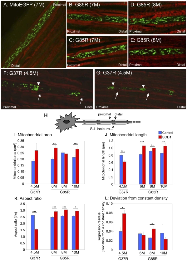
Figure 5. Mitochondria morphology is altered in mutant SOD1 axons.
MitoEGFP (green) expressed in the sciatic nerves in control (A), asymptomatic SOD1G85R 7 months (B and C), SOD1G85R 8 months (D and E), and SOD1G37R early symptomatic (F and G) labeled with Fluoromyelin red (red). Round and evenly distributed mitochondria are seen in both SOD1G85R (symptomatic stage) and SOD1G37R (early symptomatic stage). Mitochondria `pile-up or clusters are also seen in the proximal side of SLIs (arrows) in the SOD1G37R axons. Arrowheads indicate “strings” of mitochondria. Arrows indicate SLIs (H) Schematic indicating proximal and distal locations of SLIs. Mitochondrial area (I), length (J), aspect ratio (K), and density (L) were evaluated in motor axons of the sciatic nerves of and mutant SOD1 animals of various ages and age-matched MitoEGFP control animals, as described in detail in the Materials and Methods. Statistics are indicated: *, p<0.05; **, p<0.005; ***, p<0.0005.

