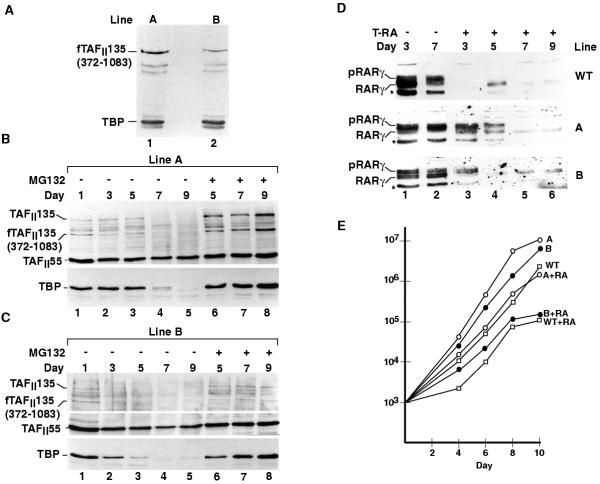
Figure 4.
Characterisation of F9 cell lines A and B expressing ectopic f TAFII135(372-1083). A. The presence of f TAFII135(372-1083) in extracts from each cell line was determined using the commercial anti-flag monoclonal antibody. B. Immunoblot analysis of extracts from line A differentiated for the number of days indicated above each. Lanes 6-8 show extracts from cells treated for 12 hours with MG132. C. Immunoblot analysis of differentiated cell extracts from line B. D. Immunoblot analysis of extracts from wild-type cells and lines A and B with antibodies against RARγ2. The native and phosphorylated species and the breakdown product are indicated as in Fig. 4D. E. Cell growth was measured for 10 days in the presence or absence of T-RA. The cell number is indicated on the X axis and the number of days of growth is indicated on the Y axis. Analogous results were obtained in three independent experiments and the graph shows the results of a representative experiment.

