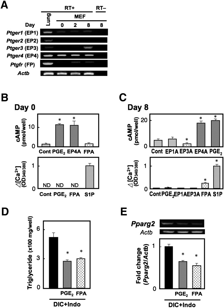
Fig. 2.
Prostanoid EP4 and FP receptors have the potential to suppress adipocyte differentiation of MEF cells. A: Gene expression of PGE and PGF receptors in MEF cells. MEF cells grown to confluency were treated with DIC, and total RNA was extracted from untreated cells (day 0), cells on day 2, or cells on day 8. Total RNA was subjected to the reverse transcription reaction in the presence (RT+) or absence of reverse transcriptase (RT−) and subsequent PCR analysis. Mouse lung RNA was used as a positive control. B, C: Undifferentiated (B, day 0) or differentiated MEF cells (C, day 8) were subjected to the cAMP (top) and Ca2+ assay (bottom). PGE2 (0.1 µM), an EP1 agonist (0.1 µM, EP1A), an EP3 agonist (0.1 µM, EP3A), an EP4 agonist (0.1 µM, EP4A), and an FP agonist (0.1 µM, FPA) were used. In the Ca2+ assay, sphingosine 1-phosphate (10 µM, S1P) was used as a positive control. D, E: MEF cells were treated with DIC supplemented with vehicle, PGE2 (1 µM), or an FP agonist (1 µM, FPA) in the presence of indomethacin (+Indo). Triglyceride content of the cells was measured on day 8 (D), and total RNA was extracted on day 8 and subjected to real time RT-PCR analysis (E). The Pparg2 gene expression levels were normalized to the β-actin (Actb) mRNA levels. Values represent the means ± SEM of three independent experiments (n = 3). *P < 0.05.

