Fig. 4.
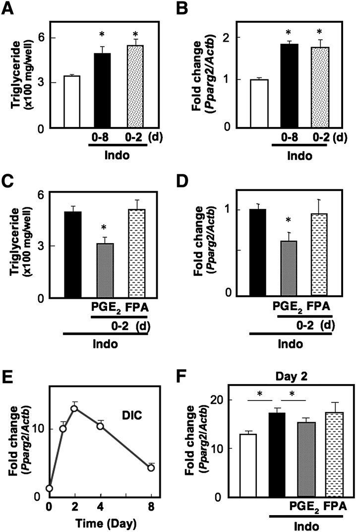
PGE2-EP4 signaling suppresses transcription of the Pparg2 gene. A, B: MEF cells grown to confluency were treated with DIC in the presence (Indo) or absence of indomethacin. On day 2, DIC was replaced with media containing insulin in the presence (0-8) or absence of indomethacin (0-2). On day 8, triglyceride content of the cells was measured (A), and Pparg2 gene expression in the cells was measured by real time RT-PCR (B). C, D: MEF cells were treated with DIC containing indomethacin (Indo) supplemented with vehicle, PGE2, or an FP agonist (FPA). On day 2, DIC was replaced with media containing insulin and indomethacin in the absence of PG receptor agonists. On day 8, triglyceride content (C) and Pparg2 gene expression (D) were measured. E: Time course of induction of Pparg2 gene transcripts in MEF cells. Cells were treated with DIC, harvested at the various time points (0 h, 9 h, day 2, day 4, and day 8) of the differentiation program, and then subjected to Pparg2 gene expression analysis. F: MEF cells were treated with DIC containing indomethacin (Indo) supplemented with vehicle, PGE2, or an FP agonist (FPA). On day 2, the cells were harvested and subjected to Pparg2 gene expression analysis. The Pparg2 gene expression levels were normalized to the β-actin (Actb) mRNA levels. Values represent the means ± SEM of three independent experiments (n = 3). *P < 0.05.

