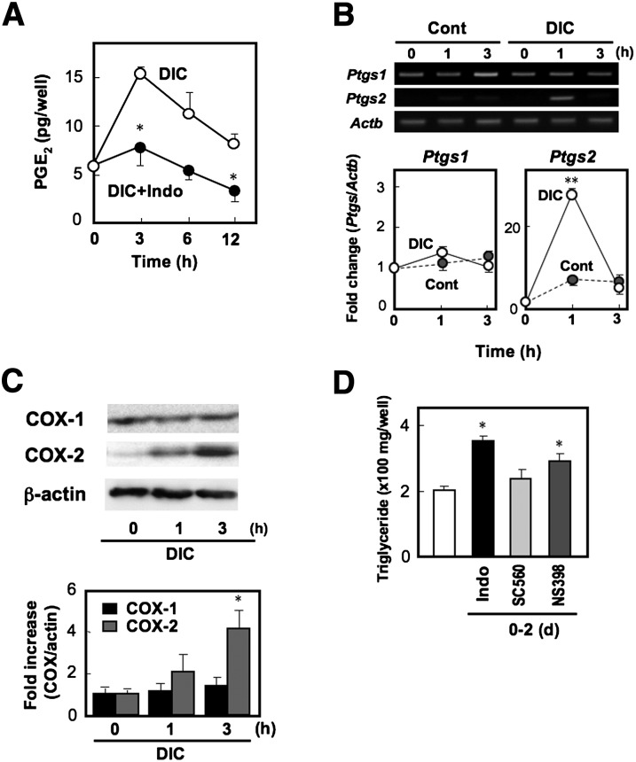
Fig. 5.
COX-2-derived PGE2 suppresses adipocyte differentiation in MEF cells. A: PGE2 production of MEF cells treated with DIC for the indicated times in the presence (+Indo) or absence of indomethacin was measured. B: MEF cells were treated with DIC or control (Cont) medium. Total RNA was isolated at the indicated times and subjected to real time RT-PCR analysis. The COX-1 (Ptgs1) and COX-2 (Ptgs2) mRNA levels were normalized to the β-actin (Actb) mRNA levels. Data are represented as a fold of the value at 0 h. C: Whole cell lysate was prepared at the indicated times and subjected to SDS-PAGE followed by immunoblotting with anti-COX-1, anti-COX-2, or anti-β-actin as a control. The histogram (bottom) shows quantitative representations of COX levels normalized to β-actin levels. D: MEF cells grown to confluency were treated with DIC supplemented with vehicle, indomethacin (Indo), COX-1 selective inhibitor SC560, or COX-2 selective inhibitor NS398 (10 µM each). On day 2, the DIC was replaced with media containing insulin without COX inhibitors. On day 8, triglyceride content was measured. Values represent the means ± SEM of three independent experiments (n = 3). *P < 0.05, **P < 0.01.

