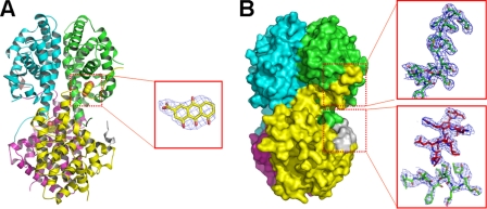
FIGURE 3.
Structure of RXRαLBD-rhein-SMRT. A, overall architecture of RXRαLBD-rhein-SMRT with the electron density of rhein (omit map contoured at 1.0σ level). B, surface of RXRαLBD-rhein-SMRT showing that the AF-2 motif of the neighboring monomer covers the ligand-binding pocket and displaces SMRT from the coregulator-binding site. The electron density map contoured at 1.0σ level shows an unambiguous positioning of the AF-2 motif (in green sticks) and SMRT corepressor motif (in red sticks).

