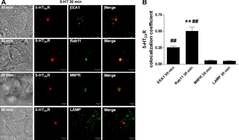
FIGURE 2.
Effects of serotonin on 5-HT2AR intracellular localization. A, serum-deprived HEK293 cells, transiently transfected with 5-HT2AR-v5 for 24 h, were incubated on ice with 1 μm 5-HT for 60 min, washed free of unbound ligand, and exposed to prewarmed ligand-free medium at 37 °C for 30 min. Cells then were fixed, stained with anti-v5 antibody (visualized with Alexa Fluor 568-conjugated secondary antibody (red)), and anti-EEA1, -Rab11, -M6PR, or -LAMP antibodies (visualized with Alexa Fluor 488-conjugated secondary antibody (green)), and analyzed by confocal microscopy. Data shown are representative of three independent experiments. Yellow indicates colocalization. White arrows pinpoint partial EEA1/5-HT2AR colocalization. The bar is 5 μm. B, the colocalizations between the 5-HT2AR-v5 and EEA1, Rab11, M6PR, or LAMP observed in panels A were quantified using Zeiss LSM 510 META colocalization analysis software. The mean colocalization coefficients, averaged from at least 16 independent single-cell images, represent pixel overlap between the 5-HT2AR-v5 and the respective markers. The coefficients varied from 0 to 1, with 0 corresponding to non-overlapping images and 1 corresponding to 100% colocalization. ## indicate a p of <0.001 versus M6PR and LAMP; ** indicate a p of <0.001 versus EEA1.

