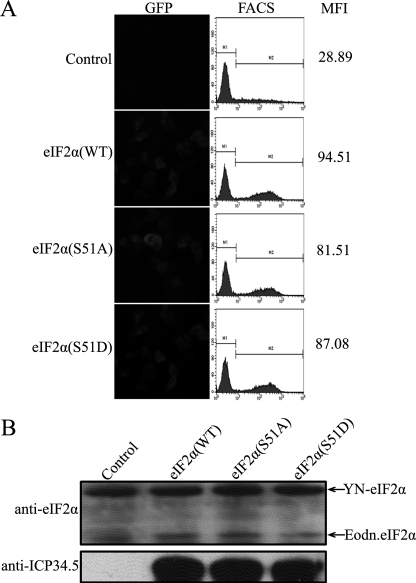
FIGURE 3.
Effect of eIF2α phosphorylation on its association with ICP34.5. A, HeLa cells were transfected with pcDNA3-YC and pcDNA3-YN-eIF2α as a control or pcDNA3-YC-ICP34.5 (WT) together with plasmids encoding YN fused to eIF2α (WT), eIF2α (S51A), or eIF2α (S51D), respectively. At 36 h post-transfection, cells were subjected to fluorescence microscopy and FACS analysis, and a FACS profile of each transfectant was plotted in the center column, with the mean YFP fluorescence intensity (MFI) in the right column. B, the protein expression levels in the BiFC assay were verified with anti-eIF2α and anti-ICP34.5 antibodies.

