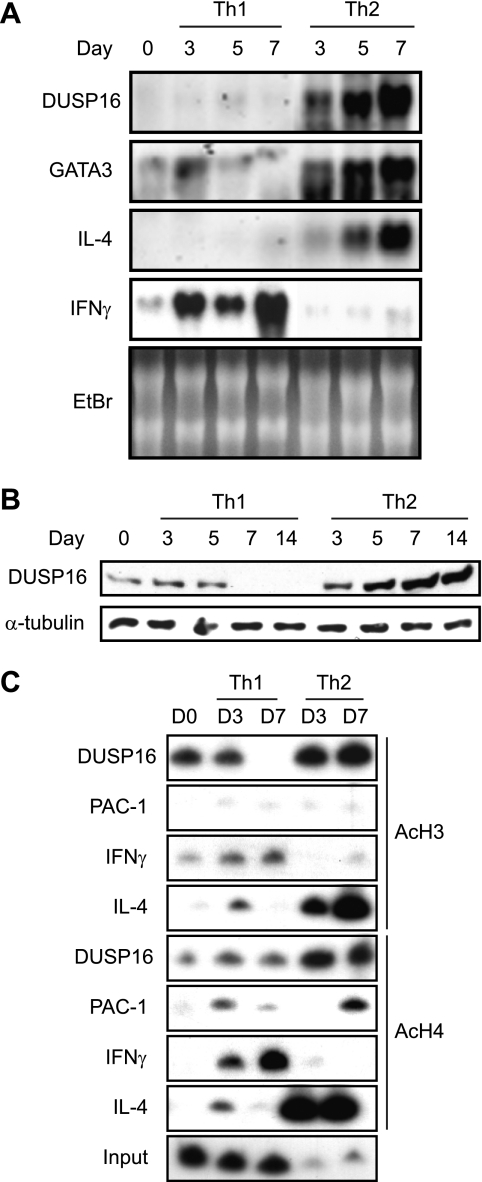
FIGURE 2.
Selective expression of DUSP16 and histone acetylation at a regulatory region of DUSP16 in differentiated Th2. A, purified CD4+ T cells were differentiated to Th1 or Th2 as described under “Experimental Procedures.” Total RNA was extracted on the indicated days, and DUSP16 gene expression was analyzed by Northern blotting using DUSP16 cDNA probe. The same blot was stripped and rehybridized with a mouse GATA-3, IL-4, or IFNγ cDNA probe. The ethidium bromide-stained gel is also shown. B, CD4+ T cells were differentiated as described in A. Total cellular lysates were prepared on the indicated days, and protein levels of DUSP16 were analyzed by Western blotting. As a control, 10% of each lysate was used to detect α-tubulin. C, purified CD4+ T cells were differentiated to Th1 or Th2 for the indicated days. Chromatin was extracted and immunoprecipitated with either anti-acetylhistone H3 or anti-acetylhistone H4 antibody. PCR analyses of DNA products for DUSP16, PAC-1, or IL-4 from immunoprecipitation reactions were carried out as described under “Experimental Procedures.”

