FIGURE 6.
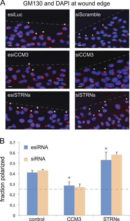
CCM3 and striatins exert opposite effects on Golgi polarization. A, GM130 and DAPI staining at the wound edge in cells depleted of indicated proteins by esiRNA (left) and chemical siRNA (right). esiLuc and siScramble are negative controls. The position of the wound is indicated by a dashed line. A 90° quadrant scoring of the Golgi positioning relative to the wound was performed (see “Experimental Procedures”). Small white asterisks indicate Golgi that are polarized toward the wound within the first cell layer. Scale bar, 10 μm. B, quantification of Golgi polarization from A indicated that ∼40% of control cells had Golgi polarized toward the wound. In cells depleted of CCM3, this value was decreased to ∼25–30%, whereas in cells depleted of striatins, this value increased to ∼54–60% (esiRNA, n = 4, p < 0.05; siRNA, n = 2). The dashed line indicates the number of cells expected to randomly orient their Golgi toward the wound in our scoring system (25%).

