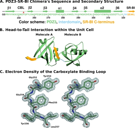
FIGURE 4.
X-ray crystal structure of the PDZ3-SR-BI target peptide chimera. A, amino acid sequence of the recombinant chimeric protein used for crystallization: N-terminal Gly-Ser dipeptide derived from the cloning vector (black), PDZ3 domain (residues 241–319 (PDZK1 numbering), green), partial interdomain segment (320–322, lies between the PDZ3 and PDZ4 domains, blue), and 5 carboxyl-terminal residues of SR-BI (505QEAKL509, SR-BI numbering, yellow). Regions of secondary structure (β strands and α helices) and the CBL are indicated above the sequence, as is the single amino acid substitution (Tyr253 → Ala) examined in this study (red). B, unit cell representation showing the head-to-tail arrangement of two PDZ3-SR-BI target peptide chimeric molecules. This figure was generated using POVScript (34) and the color scheme in panel A. C, stereo view of the electron density map (contoured at 2.2 σ) and associated molecular model of a portion of the CBL (residues 252–257) at 1.50-Å resolution.

