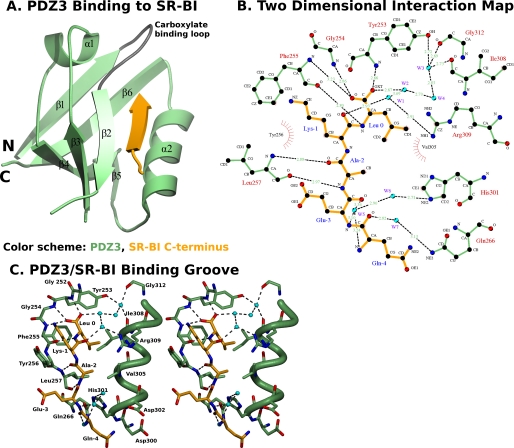
FIGURE 5.
Structure of the C-terminal SR-BI target peptide binding to PDZ3. A, ribbon diagram showing the three-dimensional structure of PDZ3 (residues 241–319, green with gray carboxylate binding loop) and the bound C terminus of SR-BI (−4QEAKL0, yellow) from an adjacent molecule. The six β-strands (β1–β6), two α-helices (α1–α2), and carboxylate binding loop (dark gray) are indicated. The vector-derived residues are not shown. B, two-dimensional representation of interactions between PDZ1 (green) and the C-terminal SR-BI target peptide (yellow). Water molecules (W1, W2, etc.) are shown as cyan spheres. Hydrogen bonds are shown as dashed lines and hydrophobic interactions as arcs with radial spokes. This figure was generated using LIGPLOT (35). C, stereo representation of the ligand-binding groove of PDZ3 (green) and the SR-BI target peptide (yellow). Oxygen atoms, nitrogen atoms, and waters are shown in red, dark blue, and cyan, respectively. Hydrogen bonds are shown as dashed lines. The orientation is similar to that in panel A.

