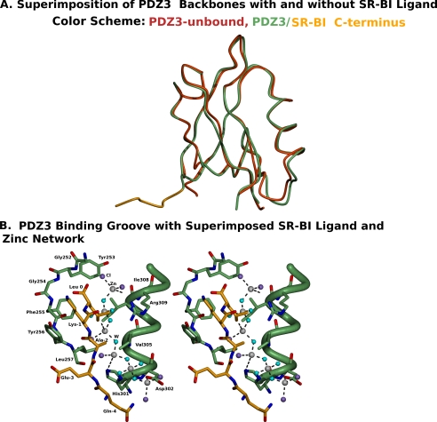
FIGURE 6.
Superimposition of the structures of PDZ3 determined with and without bound SR-BI target peptide. A, the PDZ3-SR-BI target peptide chimera is labeled green (PDZ3) and yellow (SR-BI target peptide), and the unbound PDZ3 backbone structure is labeled red. B, stereo representation of the binding groove of PDZ3 (green) and the bound SR-BI target peptide (yellow), together with the zinc-chloride-water network seen in the target peptide free PDZ3 structure (zinc, gray spheres; chloride, violet spheres; and water, cyan spheres).

