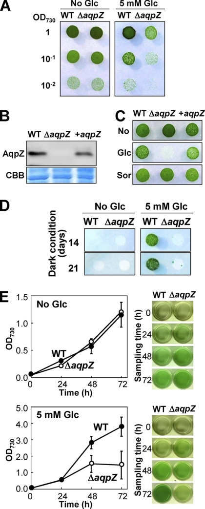
FIGURE 3.
Phenotype of the aqpZ mutants. A, WT or ΔaqpZ cells grown to mid-exponential phase were spotted onto BG11 solid medium without (left) or with 5 mm glucose (right). Each spot consisted of 5 μl of liquid culture diluted to an OD730 of 1, 0.1, or 0.01, as indicated. Plates were maintained at 30 °C, 50 μmol of photons m−2 s−1. Plates were photographed at 5 days after inoculation. B, amount of AqpZ protein in WT cells, ΔaqpZ cells, or ΔaqpZ cells complemented with the aqpZ gene (+aqpZ). Blots were probed with antiserum to AqpZ (top). Total protein (CBB) served as a loading control (bottom). C, growth of WT cells, ΔaqpZ cells, and +aqpZ cells on solid medium without an addition (top), with 5 mm glucose (middle), or with 5 mm sorbitol (bottom). D, WT or ΔaqpZ cells grown to mid-exponential phase were spotted onto solid medium without (left) or with 5 mm glucose (right). Each spot consisted of 5 μl of culture diluted to an OD730 of 1.0. The plates were incubated in the dark except for a short exposure to light (15 min each day) and photographed at days 14 and 21. E, growth curves (left) of WT (filled circles) and ΔaqpZ cells (open circles) in liquid medium without (top) and with 5 mm glucose (bottom) grown in the light. An aliquot of the cultures taken at the times indicated is shown on the right. Data and error bars (S.D.) correspond to the results of at least five independent experiments.

