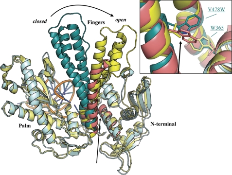
FIGURE 5.
Superposition of apo RB69 gp43 (yellow, Protein Data Bank code 1IH7) (15) and chimeric (cyan) DNA polymerase aligned on the palm domains (residues 383–468 and 573–729). The conserved region of the fingers domain of apo HSV1 (red, Protein Data Bank code 2GV9) (29) was superimposed onto the corresponding segment of apo RB69 gp43, which produced good alignment of the N-terminal helices in all three structures. The inset provides a closer view of the putative clash, marked by black arrows, between V478W and Trp-365 in the chimeric DNA polymerase. For clarity, the thumb and exonuclease domains are omitted from this diagram.

