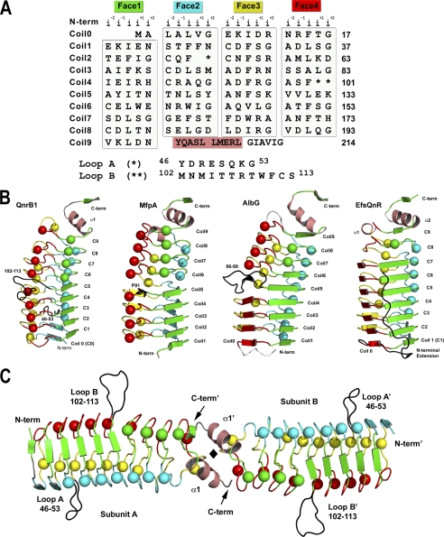
FIGURE 1.
Structure of QnrB1. A, structure-based PRP sequence diagram. The sequence of QnrB1 is segmented into four columns representing the four faces of the right-handed quadrilateral β-helix. The face name and color are represented at the top followed by the naming convention for the five residues of the pentapeptide repeats. Loops A and B are indicated by one and two asterisks, respectively, with their sequences indicated below. The N-terminal α-helix is blocked in a salmon color. B, monomer structures of QnrB1, MfpA, AlbG, and EfsQnr in a similar orientation and colored by face. C, dimeric structure of QnrB1. The A and B loops of QnrB1 are shown as a black trace. The molecular 2-fold is shown as a black diamond. Type II turn containing faces are shown as spheres, whereas type IV-containing faces are shown as strands.

